DESCRIPTION
Lactate is generated by lactate dehydrogenase (LDH) under hypoxic or anaerobic conditions. Monitoring lactate levels is, therefore, a good indicator of the balance between tissue oxygen demand and utilization and is useful when studying cellular and animal physiology. D-lactate is produced in only minor quantities in animals and measuring for Dlactate in animal samples is a means to determine the presence of bacterial infection.
Simple, direct and automation-ready procedures for measuring lactate concentration are very desirable. BioAssay Systems' EnzyChromTM lactate assay kit is based on lactate dehydrogenase catalyzed oxidation of lactate, in which the formed NADH reduces a formazan (MTT) Reagent. The intensity of the product color, measured at 565 nm, is proportionate to the lactate concentration in the sample.
APPLICATIONS
Direct Assays: D-lactate in serum, plasma, and cell media samples.
KEY FEATURES
Sensitive and accurate. Detection limit of 0.05 mM and linearity up to 2 mM D-lactate in 96-well plate assay. For cell culture samples containing phenol red: detection limit of 0.1 mM and linearity up to 1 mM D-lactate in 96-well plate assay.
Convenient. The procedure involves adding a single working reagent, and reading the optical density at time zero and at 20 min. Room temperature assay. No 37°C heater is needed.
High-throughput. Can be readily automated as a high-throughput 96- well plate assay for thousands of samples per day.
KIT CONTENTS (100 tests in 96-well plates)
Assay Buffer: 10 mL NAD Solution: 1 mL
Enzyme A: 120 µL MTT Solution: 1.5 mL
Enzyme B: 120 µL Standard: 1.0 mL 20 mM D-lactat
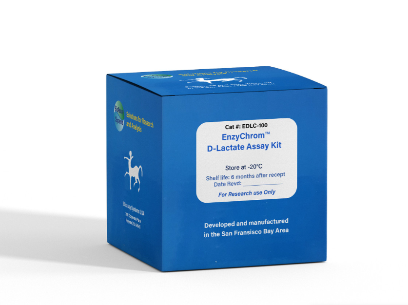
Storage conditions. The kit is shipped on ice. Store all reagents at - 20°C. Shelf life: 6 months after receipt. Precautions: reagents are for research use only. Normal precautions for laboratory reagents should be exercised while using the reagents. Please refer to Material Safety Data Sheet for detailed information.
PROCEDURES
Important: this assay is based on an enzyme-catalyzed kinetic reaction. Addition of Working Reagent should be quick and mixing should be brief but thorough. Use of a multi-channel pipettor is recommended.
1. Standard Curve. Prepare 1000 µL 2.0 mM D-lactate Premix by mixing 100 µL 20 mM Standard and 900 µL distilled water. For cell culture samples containing phenol red, prepare 1000 µL 1.0 mM lactate Premix by mixing 50 µL 20 mM Standard and 950 µL culture medium without serum. Dilute standard as follows. Transfer 20 µL standards into wells of a clear bottom 96-well plate.
Samples. Add 20 µL sample per well in separate wells. For samples with potential endogenous enzyme activity (i.e. serum, plasma,
tissue extracts), two reactions should be run: one with added Enzyme A and a No Enzyme A control. Serum and Plasma should be diluted at least 2× with dH2O prior to assay.
2. Reagent Preparation. Spin the Enzyme tubes briefly before pipetting. For each Sample and Standard well, prepare Working Reagent by mixing 60 µL Assay Buffer, 1 µL Enzyme A, 1 µL Enzyme B, 10 µL NAD and 14 µL MTT. Fresh reconstitution is recommended. For the No Enzyme A control, the Working Reagent includes 60 µL Assay Buffer, 1 µL Enzyme B, 10 µL NAD and 14 µL MTT.
3. Reaction. Add 80 µL Working Reagent per reaction well quickly. Tap plate to mix briefly and thoroughly.
4. Read optical density (OD0) for time “zero” at 565 nm (520-600nm) and OD20 after a 20-min incubation at room temperature.
5. Calculation. Subtract OD0 from OD20 for the standard and sample wells. Use the ∆OD values to determine the sample D-lactate concentration from the standard curve. For samples requiring a No Enzyme A control, subtract the ∆ODNoEnz value from the ∆ODSample and use this ∆∆OD value to determine the sample D-lactate concentration from the standard curve.
Note: if the sample OD value is higher than OD for 2 mM D-lactate standard, dilute sample in water and repeat the assay. Multiply the results by the dilution factor.
MATERIALS REQUIRED, BUT NOT PROVIDED Pipeting
(multi-channel) devices. Clear-bottom 96-well plates (e.g. Corning Costar) and plate reader.
GENERAL CONSIDERATIONS
The following substances interfere and should be avoided in sample preparation: EDTA (>0.5 mM), ascorbic acid, SDS (>0.2%), sodium azide, NP-40 (>1%) and Tween-20 (>1%).
PUBLICATIONS
[1]. Lee, JH et al (2020). Effect of cysteine on methylglyoxal-induced renal damage in mesangial cells. Cells, 9(1).
[2]. Aranda-Diaz, A et al (2020). Bacterial interspecies interactions modulate pH-mediated antibiotic tolerance. ELife, 9.
[3]. Marcu, R et al (2014). Mitochondrial Matrix Ca2 Accumulation
Regulates Cytosolic NAD/NADH Metabolism, Protein Acetylation,
and Sirtuin Expression. Mol Cell Biol. 34(15):2890-902.
